
Where are the kidneys?
Most people have two kidneys, which sit at the back of the abdomen (behind the liver and intestines) in the small of the back on either side of the spine. Each is 11-14 cm (5-6 inches) long and is bean-shaped. It is very difficult to examine a normal kidney because they are far back when examined from the front, and behind a lot of muscles when examined from the back. From the kidneys, the collecting systems funnel the urine into tubes (ureters) which go down to the bladder. The connection to the outside is along the urethra.
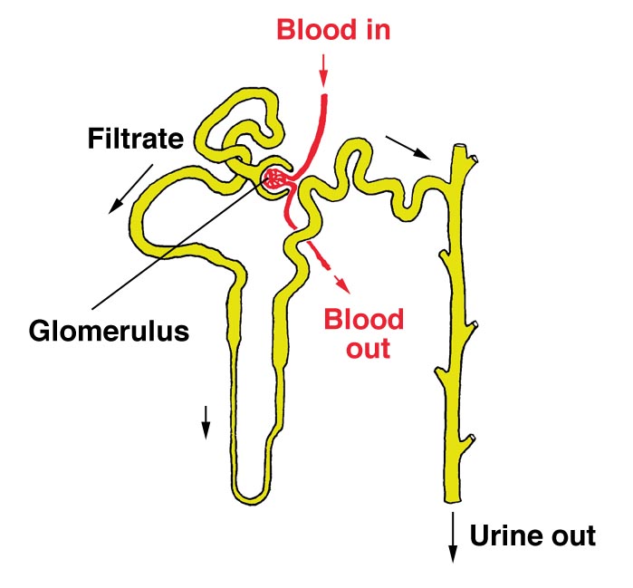
Each kidney is joined to the largest artery in the body, the aorta, by a short renal artery. The kidneys have a huge blood supply – one fifth (20%) of normal blood flow leaving the heart.
ANIMATION
Click here to see an animation of where the kidneys are, and a bit about how they work, from the
Kidney Patient Guide (Wrexham Maelor Hospital, Wales). You need Flash Player on your PC (or Mac) to see it work (links to get this free are provided).
What do the kidneys do?
- remove toxic waste products
- remove excess water and salts
- play a part in controlling your blood pressure
- produce erythropoietin (epo for short) which stimulates red cell production from the bone marrow – you get anaemic without this
- help to keep calcium and phosphate in balance for healthy bones
- maintain the blood in a neutral (non-acid) state
Signs that the kidneys are not working properly
Urine tests may show blood or protein that should not normally be there. More information on haematuria (blood in the urine) and proteinuria (protein in the urine) is given on other pages.
Blood pressure may be high. Most people with high blood pressure do not have serious kidney disease, but high blood pressure can be a sign of kidney disease. It is more likely to be connected to kidney disease in people who are young, or have severe high blood pressure.
Trouble passing urine is rarely caused by kidney trouble – unless the kidneys are very severely damaged. It is more commonly caused by problems in the bladder, or in the nerves supplying the bladder, or by infection in the urine.
Pain around the kidneys is an uncommon symptom in kidney disease except with kidney stones, and usually has alternative explanations. It is very common with kidney stones, and sometimes occurs with urine infections. Patients with large cysts in the kidney may get pain from them. Sometimes blockage of the artery to the kidney causes pain. Otherwise pain is unusual in kidney disease.
Other symptoms come from loss of kidney function. In the early stages of many kidney diseases, there are no symptoms at all. Kidney function needs to be quite badly damaged before any symptoms become noticeable. At first these are usually very vague and non-specific, and easily confused with many other conditions. A general slowing down and tiredness are common. Later symptoms may include loss of appetite, itch, poor sleep, and many others. Some of these are described on our pages on chronic renal failure and its progression.
Tests for kidney disease
| Blood tests (more info about individual kidney blood tests) | |
| Urea | a simple test, but the result is affected by food and by dehydration. |
| Creatinine | a more reliable measure. |
| Estimating GFR using just blood tests (eGFR, estimated GFR) | |
| Equations | The MDRD and CKD-EPI equations use blood tests, age, and race to work out approximately what the GFR is. They are not as good as measuring GFR directly, but they are very useful. Calculate your MDRD eGFR (from the Renal Association website); or your CKD-EPI eGFR (Kidney Health Australia) Remember that this is not as accurate as measuring GFR, and in some people eGFR may be quite far from the real GFR. Explain reduced GFR |
| More accurate tests to show GFR GFR means glomerular filtration rate, the most useful measure of kidney function. As the normal value is about 100, it gives an approximate ‘score out of 100’ for kidney function. However your normal GFR depends on your size and age. |
|
| Creatinine clearance | requires collection of all urine over 24h, with a blood test. The Cockroft-Gault equation used to approximately estimate this before eGFR equation. |
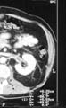
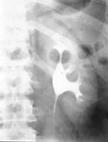
Imaging tests (X-rays etc.)
A number of different kinds of test can be used to show the kidneys. These are some common examples.

Kidney biopsy
A kidney biopsy involves taking a small sample of kidney through a needle to look at it under the microscope. More information about this test is given in our page on renal biopsy.
Where can I find further information?
You can search this site, or go to our EdRen INFO page. Or you can brave the rest of the Internet.
Acknowledgements: The author of this page was Neil Turner. It was first published in January 2000, with revisions in February 2016. The date is was last modified is shown in the footer.